
PET/CT Examination
-
 0
0
-

-

-


What is Positron Emission Tomography – Computed Tomography (PET/CT) Scanning?
Positron emission tomography, also called PET imaging or a PET scan, is a kind of nuclear medicine imaging. PET scan measures important body functions, such as blood flow, oxygen use, and sugar (glucose) metabolism to help doctors evaluate how well organs and tissues are functioning.
CT imaging uses special x-ray equipment, and in some cases a contrast material, to produce multiple images or pictures of the inside of the body. These images can then be interpreted by a radiologist on a computer monitor. CT imaging provides excellent anatomic information.
Today, almost all PET scans are performed on instruments that are combined PET and CT scanners. The combined PET/CT scans provide images that pinpoint the anatomic location of abnormal metabolic activity within the body. The combined scans have been shown to provide more accurate diagnoses than the two scans performed separately.
As a most high-end medical imaging devise in the world, PET/CT is currently the only technology in anatomic form to obtain images of function, metabolism.
Featuring no wounds, it detects the minimum cancer focus and discovers potential cancerous focus early of high resolution and sensitivity. With accurate diagnostic rate of more than 90%, PET/CT plays an important role in planning treatments.
What are some common uses of the PET/CT?
PET and PET/CT scans are performed to:
1. detect cancer.
2. determine whether a cancer has spread in the body.
3. assess the effectiveness of a treatment plan, such as cancer therapy.
4. determine if a cancer has returned after treatment.
5. determine blood flow to the heart muscle.
6. determine the effects of a heart attack, or myocardial infarction, on areas of the heart.
7. identify areas of the heart muscle that would benefit from a procedure such as angioplasty or coronary artery bypass surgery (in combination with a myocardial perfusion scan).
8. evaluate brain abnormalities, such as tumors, memory disorders, seizures and other central nervous system disorders.
9. map normal human brain and heart function.
>>For more knowledge about cancer treatment technology, please click online doctors for consultation.
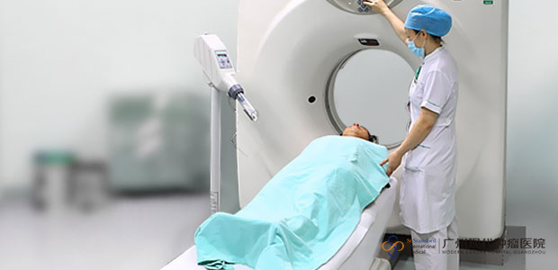
What does the equipment look like?
A PET scanner is a large machine with a round, doughnut shaped hole in the middle, similar to a CT or MRI unit. Within this machine are multiple rings of detectors that record the emission of energy from the radiotracer in your body.
The CT scanner is typically a large, box-like machine with a hole, or short tunnel, in the center. You will lie on a narrow examination table that slides into and out of this tunnel. Rotating around you, the x-ray tube and electronic x-ray detectors are located opposite each other in a ring, called a gantry. The computer workstation that processes the imaging information is located in a separate control room, where the technologist operates the scanner and monitors your examination in direct visual contact and usually with the ability to hear and talk to you with the use of a speaker and microphone.
Combined PET/CT scanners are combinations of both scanners and look similar to both the PET and CT scanners.
What are the benefits of PET-CT ?
Nuclear medicine examinations offer information that is unique—including details on both function and structure—and often unattainable using other imaging procedures. For many diseases, nuclear medicine scans yield the most useful information needed to make a diagnosis or to determine appropriate treatment, if any. Nuclear medicine is less expensive and may yield more precise information than exploratory surgery. By identifying changes in the body at the cellular level, PET imaging may detect the early onset of disease before it is evident on other imaging tests such as CT or MRI.
The benefits of a combined PET/CT scanner include:
1. greater detail with a higher level of accuracy; because both scans are performed at one time without the patient having to change positions, there is less room for error.
2. Greater convenience for the patient who undergoes two exams (CT & PET) at one sitting, rather than at two different times.
>>For more knowledge about cancer treatment technology, please click online doctors for consultation.
How does the PET/CT work?
With ordinary x-ray examinations, an image is made by passing x-rays through the body from an outside source. In contrast, nuclear medicine procedures use a radioactive material called a radiopharmaceutical or radiotracer, which is injected into your bloodstream, swallowed or inhaled as a gas. This radioactive material accumulates in the organ or area of your body being examined, where it gives off a small amount of energy in the form of gamma rays. A gamma camera, PET scanner, or probe detects this energy and with the help of a computer creates pictures offering details on both the structure and function of organs and tissues in your body.
Unlike other imaging techniques, nuclear medicine imaging exams focus on depicting physiologic processes within the body, such as rates of metabolism or levels of various other chemical activity, instead of showing anatomy and structure. Areas of greater intensity, called "hot spots," indicate where large amounts of the radiotracer have accumulated and where there is a high level of chemical or metabolic activity. Less intense areas, or "cold spots," indicate a smaller concentration of radiotracer and less chemical activity.
What is the Procedures for PET-CT?
Before the exam, the patient fasts and forbids glucose transfusion for greater than 6 hours; meanwhile avoids doing strenuous or long-time exercise. Nurse introduces exam procedures, measures patient’s height and weight, and then finishes the exam result.Doctor talks to patient about his/her medical history, checks patent’s blood glucose, explains to patient about informed consent before patient or his/her family sign it.
Patient takes a rest for 45 minutes after PDG injection.Patient keeps quiet, walks less and avoids talking to others as possible; he / she also have to urine completely before CET/PT.
PET-CT takes 30 minutes.
In order to finish the PET-CT for whole-body, 2.5-3 hours should be allowed in advance.
>>For more knowledge about cancer treatment technology, please click online doctors for consultation.
Patient Stories
-
 Ovarian cancer
- Winda Fitrianah Muchlis
- Indonesia
- Treatment programs: Interventional therapy
- Description: I am Winda Fitrianah Muchlis from Jakarta, Indonesia, currently 45 years old. Now that I am rosy-cheeked, can you imagine that I am a patient who was once diagnosed with stage IV ovarian cancer?
- more >
-
 other
Ewing's sarcoma
other
Ewing's sarcoma
- Haten
- Treatment programs: interventional therapy + radical surgery
- Description: Haten is a lively 22-year-old teenager who enjoys sports, particularly playing basketball. He recently started his career and was looking forward to a bright future. Unfortunately, in June 2023, Haten received the devastating news that he had been diagnosed with Ewing's sarcoma, which is a malignant tumor that affects the bones.
- more >
-
 Breast cancer
- Elisabeth
- Netherlands
- Treatment programs: Interventional therapy + natural therapy
- Description: Many desperate cancer patients are declared untreatable by local doctors, but you will see hope here. The hospital focuses on minimally invasive techniques and the patient is treated at the center. Don't waste too much time in hesitation and stumbling, you need timely treatment and you have more options!" This is my heartfelt appeal to cancer patients.
- more >
-
 other
other
- Lukman Holy
- Indonesia
- Treatment programs: Interventional therapy
- Description: "I am glad to have found St. Stamford Modern Cancer Hospital Guangzhou because the minimally invasive treatment technology here does not harm normal tissues of the body. Compared with the pain caused by the side effects of systemic chemotherapy, I am really lucky! During the treatment of cancer, I can still maintain a good quality of life, which is a blessing brought by the new technology of cancer treatment. "This is my true experience after treatment at St. Stamford Modern Cancer Hospital Guangzhou. On January 11, 2024, I came to the hospital for further treatment and was interviewed by the hospital After the first systemic chemotherapy, I fainted and fell to the ground I am Lukman Holy from Makassar, Indonesia. In 2022, I started to feel unwell: gum discomfort, frequent runny nose, and the runny nose was accompanied by bloodshot eyes! I felt uneasy at that time and had a premonition: something big must have happened to me! I was rushed to the local hospital for examination.
- more >
-

- Tan Tanty Bintari
- Indonesia
- Treatment programs: Urachal adenocarcinoma
- Description: Tan Tanty Bintari from Surabaya, Indonesia, was diagnosed with stage IV urethral adenocarcinoma due to blood in her urine, with a tumor size of about 3.8cmx4.4cmx2.2cm and multiple lung metastases, and a tumor size of about 2.4x1.8cm in her left lung. After comprehensive minimally invasive treatment at St. Stamford Modern Cancer Hospital Guangzhou, the tumor has almost disappeared and her life quality is good now.
- more >
-
 Colon cancer
- Jefferies
- Indonesia
- Treatment programs: Interventional therapy + Cryotherapy
- Description: Jeffery from Indonesia, 54 years old, was diagnosed with colon cancer more than a year ago. To seek a more suitable treatment method for himself, he learned about minimally invasive treatment through the Internet. He came to St. Stamford Modern Cancer
- more >
